UF-led study could lead to new way to detect brain changes associated with Alzheimer’s risk
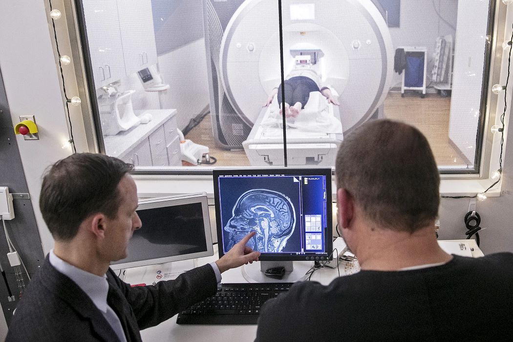
David Vaillancourt, Ph.D., (left) is a senior author on a new study linking abnormal amyloid in the blood with brain changes on diffusion MRI.
New research published today suggests there is a link between abnormal blood levels of amyloid — a protein associated with Alzheimer’s disease — and subtle changes in brain microstructures on a type of MRI, findings that could lead to a new way to detect Alzheimer’s earlier in people with no clinical signs.
Researchers analyzed the results of 128 human participants with and without dementia from the 1Florida Alzheimer’s Disease Research Center who underwent imaging scans using an established diagnostic tool called positron emission tomography, or PET, which can detect amyloid plaques in the brain, a hallmark of Alzheimer’s disease.
Even when a PET scan was negative for amyloid and a participant free of dementia symptoms, researchers found there was an association in those who showed abnormal amyloid levels in the blood and structural abnormalities in the brain detected through a newer method called diffusion MRI, also known as “free-water” imaging.
A team led by investigators from UF's Evelyn F. and William L. McKnight Brain Institute and the Norman Fixel Institute for Neurological Diseases at UF Health reported that the results represent a novel finding that free-water imaging is sensitive to early stages of decline in brain tissue and tiny structures in key parts of the brain — even when a PET scan is negative. The results were published in Alzheimer’s & Dementia: The Journal of the Alzheimer’s Association.